Beware the foot sprain
Case of a missed Lisfranc injury.
A 66-year-old man presented with persistent swelling and pain in his left foot after a trip and fall at a bus stop. At his first clinic visit he was able to partially weight-bear. Examination documented tenderness over the midfoot and base of the first metatarsal, but no plantar bruising was recorded. An X-ray was initially interpreted as normal and formally reported as showing no fracture or dislocation, so the injury was treated as a sprain.
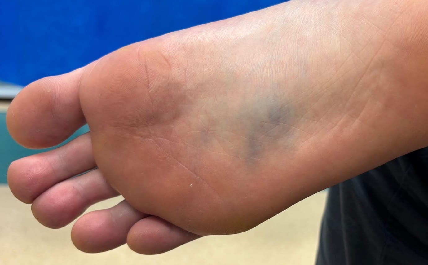
One week later he returned with worsening pain, swelling, and the sensation that “something was moving” inside his foot. On examination there was significant swelling over the dorsum, bruising on the lateral and plantar aspects, and marked midfoot tenderness. Circulation and sensation were intact, but the severity of symptoms seemed out of proportion to a simple sprain, raising suspicion of a fracture or Lisfranc injury.
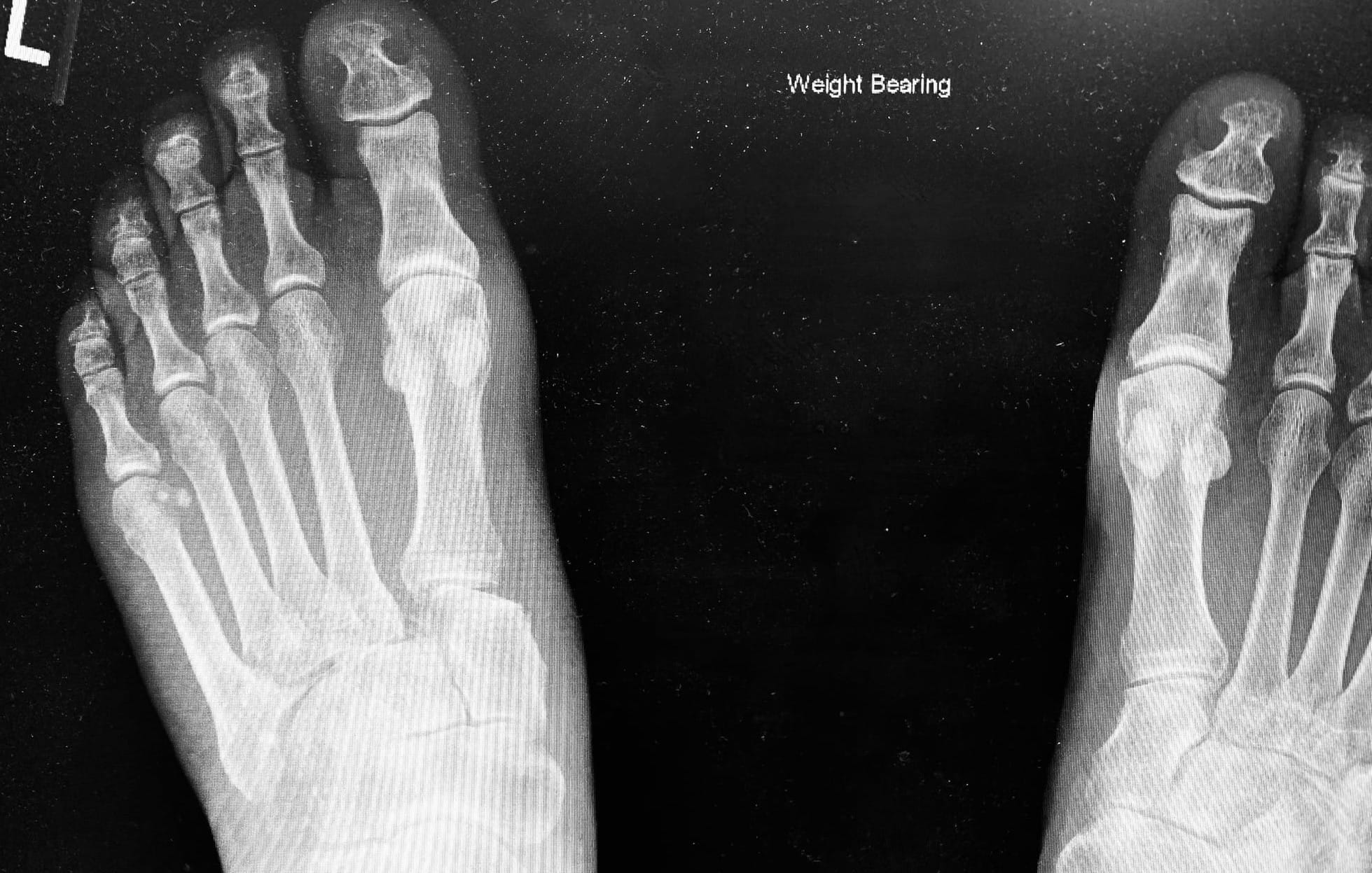
A repeat X-ray confirmed the diagnosis. There was gross subluxation of the first and second tarsometatarsal joints with 6–7 mm displacement, and a possible cuboid fracture. The patient was urgently referred to orthopaedics for open reduction and internal fixation.
This case illustrates the difficulties in the diagnosis of Lisfranc injury. Some studies suggest that 20-40% of Lisfranc injuries are missed at initial presentation.1,2 The patient was initially seen by a junior member of staff. In the clinical notes there was no record of plantar bruising or provocative tests. Good safety netting occurred though and the patient re-presented.
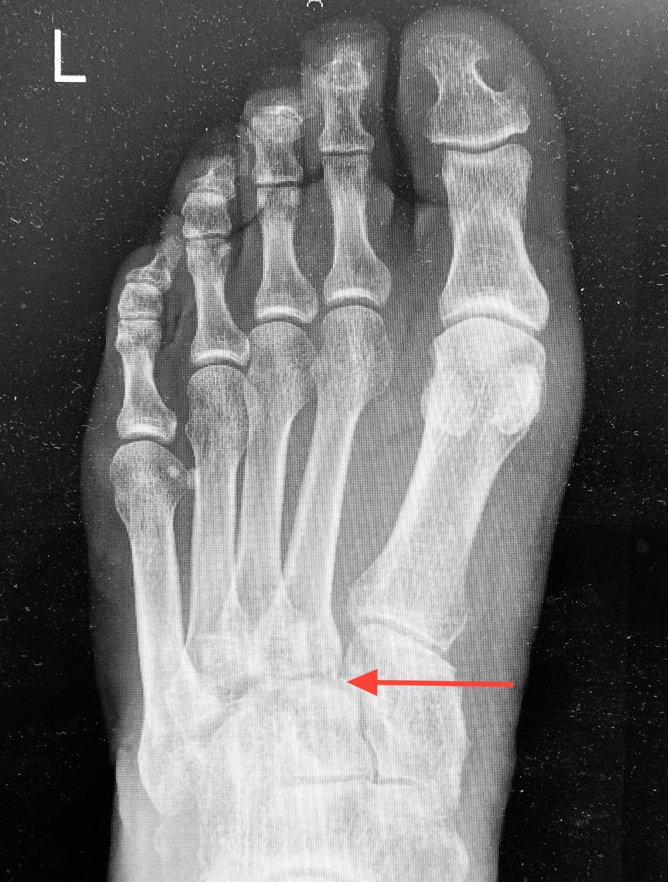
The initial x-ray was reported normal by a consultant radiologist. The size of the first inter- metatarsal space was suspicious and on closer review there was a subtle abnormality; a small step at the junction of the base of the second metatarsal with the middle cuneiform(see arrow).
The high, ongoing misdiagnosis rate of Lisfranc injuries would tend to suggest that there is probably an under recognition of many of the subtleties of clinical examination/provocative tests and radiology of Lisfranc injuries and this is what teaching should perhaps focus on. Delayed diagnosis can lead to significant morbidity and early diagnosis is essential.1,2
Learning nuggets
*Lisfranc injuries can be due to high or low energy mechanisms of injury; the latter can be misleading.1,2
*Provocative tests include: piano-key, mid-foot compression, compressions across the width of the foot, abduction-pronation.1,2 It is unclear from the literature how useful these tests are.
*Plantar bruising has been considered pathognomonic but studies show it is only present in about 50 % cases.3
*There needs to be a high index of suspicion to avoid missing the diagnosis especially in children.3
*Arterial injury and compartment syndrome are rare but emergency complications of Lisfranc injury.They are more associated with high-energy mechanisms. Compartment syndrome may occur in 25 % of cases.4
*Non-weight bearing x-rays miss subtle diastasis between the 1st and 2nd metatarsal bases in up to 50 % cases.5
*The fleck sign is traditionally considered pathognomonic of Lisfranc injury but overall incidence is difficult to confirm.
*Bilateral weight-bearing views detect about 85% of injuries and should be requested if there is any suspicion of possible Lisfranc injury.5
*Other key subtle X-ray changes to look for are (a) malalignment of the 2nd tarsometatarsal joint and (b) a dorsal step on the lateral view due to dorsal displacement of the 2nd metatarsal base.6
*One study showed CT and MRI have a sensitivity of 72.2 and 87.0 respectively.5 If the patient is unable to comply with weight-bearing views and there is a high index of suspicion an urgent CT could be requested from the local orthopaedic team.
References
1. Royal Australian College of General Practitioners. Lisfranc Injuries. Available from: https://www.racgp.org.au/afp/2017/march/lisfranc-injuries
2. Moracia-Ochagavía I, Rodríguez-Merchán EC. Lisfranc fracture-dislocations: current management. EFORT Open Reviews. 2019;4(7):430–44. Available from: https://eor.bioscientifica.com/view/journals/eor/4/7/2058-5241.4.180076.xml
3. Kushare I, Wunderlich N, Elabd A, Attia E. Pediatric and adolescent Lisfranc injuries — Presentation, treatment and outcomes. The Foot. 2021;46:101737. Available from: https://linkinghub.elsevier.com/retrieve/pii/S0958259220300754
4. AO Foundation. Treatment of Lisfranc Injuries. Available from: https://surgeryreference.aofoundation.org/orthopedic-trauma/adult-trauma/metatarsals/lisfranc-injury/treatment-of-lisfranc-injuries
5. Tang L, Zhou W, Bai L, Wu C, Xiong C, Yan Y, et al. Comparison of diagnostic performance of X-ray, CT and MRI in patients with surgically confirmed subtle Lisfranc injuries. Exp Ther Med. 2024;27(4):174. Available from: http://www.spandidos-publications.com/10.3892/etm.2024.12462
6. Knipe H, Gaillard F. Lisfranc injury. Radiopaedia.org. Available from: http://radiopaedia.org/articles/1590