Paradoxical Findings
A sudden visual field defect after limb trauma.

I was on duty one weekend as a Medical Registrar at a base hospital in NZ. I had a call from the duty Orthopaedic consultant asking for a medical consult for an patient on the orthopaedic ward. The patient was a man in his sixties who had been admitted the day before with a degloving injury of the lower leg during an MVA. The patient had described some sudden visual loss on the ward and the Orthopaedic consultant had diagnosed a homonymous hemianopia. I agreed. I put the phone down and admittedly had a small chuckle to myself at the thought of a Orthopaedic consultant diagnosing a visual field defect.
The plot thickened. The patient turned out to be the father-in-law of my house officer at the time— so no pressure.
To my great surprise the Orthopaedic consultant was absolutely right! The patient had an isolated homonymous hemianopia. There were no other neurological signs. He was well looking with GCS15. The patient confirmed that this had developed overnight and was not present prior to the accident. This is strange I thought.
I requested a CT Brain which confirmed an occipital cerebral infarct. I went back to review the patient. He was in sinus rhythm and had normal heart sounds and an unremarkable ECG. This was bugging me— why would he suddenly have an occipital infarct after an episode of trauma?
As I was considering the possibilities the patient then causally mentioned that he had also had some vague right upper chest. Chest exam and oxygen saturation were normal. I looked down at the injured leg. It was very swollen. I asked the Orthopaedic consultant about the likelihood of an underlying DVT in the affected leg. He thought almost certainly.
General Internal Medicine is a specialty which requires you to always think broadly and at the same time make connections between seemingly unconnected things. I started wondering if this patient had sustained a paradoxical embolus (PDE). I had never seen one before and only read about it for the MRCP exam.
A venogram confirmed an extensive above knee DVT. A CTPA confirmed a pulmonary embolus. This was a perfect scenario for a PDE.
I discussed the situation with my consultant who was a neurologist. With the obvious anticoagulation dilemma aside he could not believe that a venous embolus could pass through an intra-cardiac shunt and then into a small vertebral artery to cause an occipital infarct. He admitted though that we should try to exclude a patent foramen ovale (PFO).
I discussed the case with the hospital cardiologist who reluctantly agreed to perform a trans-oesophageal echo. It was a difficult procedure which had to be terminated prematurely. We did not demonstrate a PFO but to this day I am skeptical as to the accuracy of the test. The cardiologist did not want to investigate things further. I have remained convinced that this patient had a PDE.
Learning Nuggets
- A paradoxical embolism occurs when thromboembolic material originating in the venous circulation crosses an intracardiac or pulmonary right-to-left shunt and enters the systemic arterial circulation. The classic scenario is a DVT passing through a PFO and subsequently embolising to the brain, coronary circulation, viscera or a limb.1
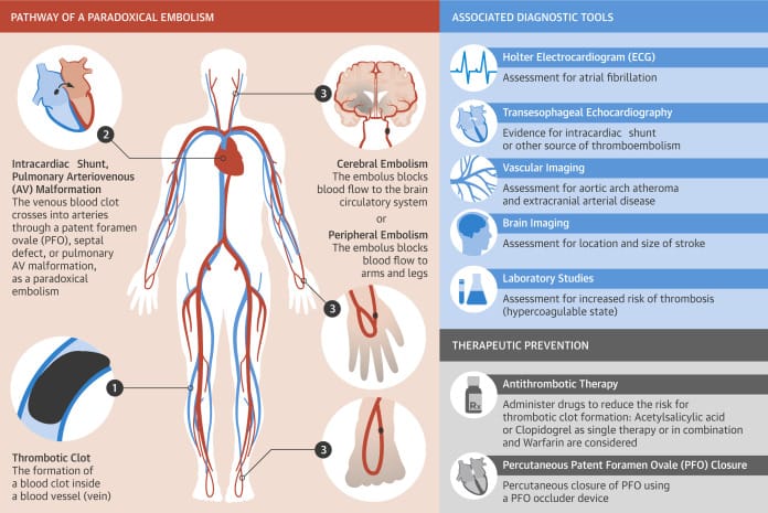
- The clinical effects of embolisation can be varied. Most commonly ischaemic stroke but MI, mesenteric ischaemia, renal infarction and peripheral limb ischaemia have been described.1
- The true prevalence of PDE is difficult to know due to the diagnostic difficulty and under reporting.1,2
- Emboli can be blood clot, air, fat particles, amniotic fluid or tumour.2
- Patent foramen ovale (PFO) is the most commonly associated intracardiac shunt and is present in about 25-30% of the population.1 Left atrial pressure is usually higher than right atrial pressure. Elevated right atrial pressure can occur and reverse the shunt. This can occur with pulmonary embolus or transiently by Valsalva type maneouvers such as coughing, sneezing, squatting, defecation and micturition.1
- Cerebral infarction is the most common manifestation of PDE. Up to 45% of strokes are cryptogenic. Cryptogenic stroke patients have a more than 2-fold prevalence of PFO compared to those with conventional strokes.1
- The prevalence of intracardiac right-to-left shunts in patients with migraine has been reported to be up to 50%. Paradoxical small embolisation has been suggested as a cause of migraine.1
- A 2024 systematic review and meta-analysis showed PFO was significantly associated with ischaemic stroke in patients with PE compared to those without PFO (odds ratio 5.36, 95 % CI 3.20-8.99).3
- The 2024 European Stroke Organisation (ESO) Guidelines on the diagnosis and management of patent foramen ovale (PFO) after stroke recommends PFO closure for patients aged 18–60 years who do not have any other apparent reason for their stroke except the PFO.4
- The 2022 SCAI Guidelines for the Management of Patent Foramen Ovale present a number of recommendations related to other scenarios.4
- In migraine patients without a prior PFO-associated stroke, the guideline panel suggests against the routine use of PFO closure for the treatment of migraine.4
- In SCUBA divers with prior decompression illness and without a prior PFO-associated stroke, the guideline panel suggests against the routine use of PFO closure to prevent DCI.4
- In patients with documented thrombophilia and without a prior PFO-associated stroke, the guideline panel suggests against the use PFO closure in addition to antithrombotic therapy.4
- In persons with systemic embolism and without a prior PFO-associated stroke, in whom other embolic etiologies have been excluded, the guideline panel suggests PFO closure rather than medical therapy alone.4
Further Reading
1. Windecker S, Stortecky S, Meier B. Paradoxical Embolism. Journal of the American College of Cardiology [Internet]. 2014; 64(4):403–15. Available from: https://linkinghub.elsevier.com/retrieve/pii/S0735109714028447.
2. Hakman EN, Cowling KM. Paradoxical Embolism. In: StatPearls [Internet]. Treasure Island (FL): StatPearls Publishing; 2026. Available from: http://www.ncbi.nlm.nih.gov/books/NBK470196/.
3. Lucas TO, Schaustz EB, Dos Reis IJR, Lopes CG, Mendoça VS, Salluh JIF, et al. Risk of ischemic stroke in patients with pulmonary embolism and patent foramen ovale: A systematic review and meta-analysis. Journal of Stroke and Cerebrovascular Diseases [Internet]. 2025; 34(1):108157. Available from: https://linkinghub.elsevier.com/retrieve/pii/S1052305724006001.
4. Caso V, Turc G, Abdul-Rahim AH, Castro P, Hussain S, Lal A, et al. European Stroke Organisation (ESO) Guidelines on the diagnosis and management of patent foramen ovale (PFO) after stroke. European Stroke Journal [Internet]. 2024; 9(4):800–34. Available from: https://academic.oup.com/esj/article/9/4/800/8269146.
5. Kavinsky CJ, Szerlip M, Goldsweig AM, Amin Z, Boudoulas KD, Carroll JD, et al. SCAI Guidelines for the Management of Patent Foramen Ovale. Journal of the Society for Cardiovascular Angiography & Interventions [Internet]. 2022; 1(4):100039. Available from: https://linkinghub.elsevier.com/retrieve/pii/S2772930322000230.