The Great Pretender Revisited
Ten Rules for Ectopic Pregnancy

I sometimes look back on my career and think about the experiences that have shaped me most as a doctor. One of those occurred during my final year of medical school.
I was on my gynaecology attachment at a London teaching hospital, shadowing the duty registrar. One evening we were called to the Emergency Department to assess a woman in her thirties who presented with vaginal bleeding and a positive pregnancy test. Her observations were normal. She looked entirely well, sitting comfortably upright on a trolley in an examination cubicle.
The registrar had only just begun taking her history when the patient quietly said she wasn’t feeling well. Within moments she became pale and clammy. Her level of consciousness deteriorated. We laid her flat. She became unresponsive, then started groaning with severe abdominal pain as her blood pressure plummeted.
At exactly the same time, a patient in cardiac arrest arrived in the Emergency Department, and the entire ED team disappeared into the resuscitation room. The gynaecology registrar suddenly found herself managing a critically ill patient with only a final-year medical student for assistance.
We inserted intravenous lines and began fluid resuscitation. The registrar diagnosed a ruptured ectopic pregnancy. There was no bedside ultrasound in those days. We transferred the patient immediately to theatre, running through the hospital corridors while I squeezed bags of intravenous fluid. Surgery confirmed a ruptured tubal ectopic pregnancy with a large haemoperitoneum. Fortunately, she survived.
Although I was only a medical student, that experience has stayed with me ever since. What struck me most was how a young woman who appeared completely well could deteriorate so catastrophically in a matter of minutes.
Over the years I have often encountered what I consider an overly relaxed attitude towards ectopic pregnancy from healthcare professionals across many disciplines. Many have never witnessed an ectopic pregnancy “crash” before their eyes. I have also found that systems of care—particularly access to urgent ultrasound—are often insufficiently responsive to the potential urgency of the condition. More than once I have found myself frustrated trying to convey just how dangerous ectopic pregnancy can be.
My experience as a medical student gave me an “ectopic paranoia” which has stayed with me ever since. I still feel fortunate though that I have never missed the diagnosis.
I have managed many ectopic pregnancies during my career, but a few cases remain particularly memorable.
I was working in an urgent care clinic in a ski resort when a ski instructor came down from the mountain at the end of the day complaining of mild lower abdominal pain and a small amount of vaginal bleeding. She looked entirely well. Her vitals were normal. She was not even tachycardic. Her abdomen was soft and non-tender.
The nurse informed me that her urine β-hCG was positive. Purely by chance that day I was able to arrange an immediate on site ultrasound . It demonstrated an adnexal mass with a large volume of free fluid in the pelvis.
I explained that she likely had a ruptured ectopic pregnancy and required emergency transfer by helicopter to the base hospital. She could not understand what all the fuss was about and wanted to go home first to collect some belongings. She did not understand that the seriousness of the situation. I insisted that she not leave the clinic. Reluctantly, she agreed and allowed me to insert two large-bore IV cannulae.
On another occasion, a colleague in general practice saw a woman in early pregnancy with light vaginal bleeding. The bleeding was diagnosed as implantation bleeding, and no ultrasound was requested. When an ectopic pregnancy was eventually diagnosed, my colleague complained that she had been given the wrong gestational dates.
A staff member at another clinic where I worked mentioned to the senior nurse that she had developed some lower abdominal pain. To the nurse’s credit, she immediately performed a urine pregnancy test, which was positive. The patient looked entirely well and had normal observations. We arranged an urgent ultrasound on site. Once again, the scan showed an adnexal mass with a significant pelvic collection of blood. Once again, the patient appeared comfortable, was haemodynamically stable, and gave little outward indication of the seriousness of her condition.
The final case occurred late on a Friday afternoon when I was a senior registrar Emergency Medicine. I felt that a patient required ectopic pregnancy to be excluded. I performed a point-of-care ultrasound, which was unremarkable. The duty radiology registrar initially told me that it was too late in the day for a formal scan and that the patient should return on Monday! The gynaecology service at that hospital was not known for readily admitting patients, and I was not optimistic that they would help. Fortunately, at the last minute, a formal ultrasound was performed. An ectopic pregnancy was confirmed.
Even though there were no adverse outcomes I would still class these cases as near-misses. The outcomes could have been very different.
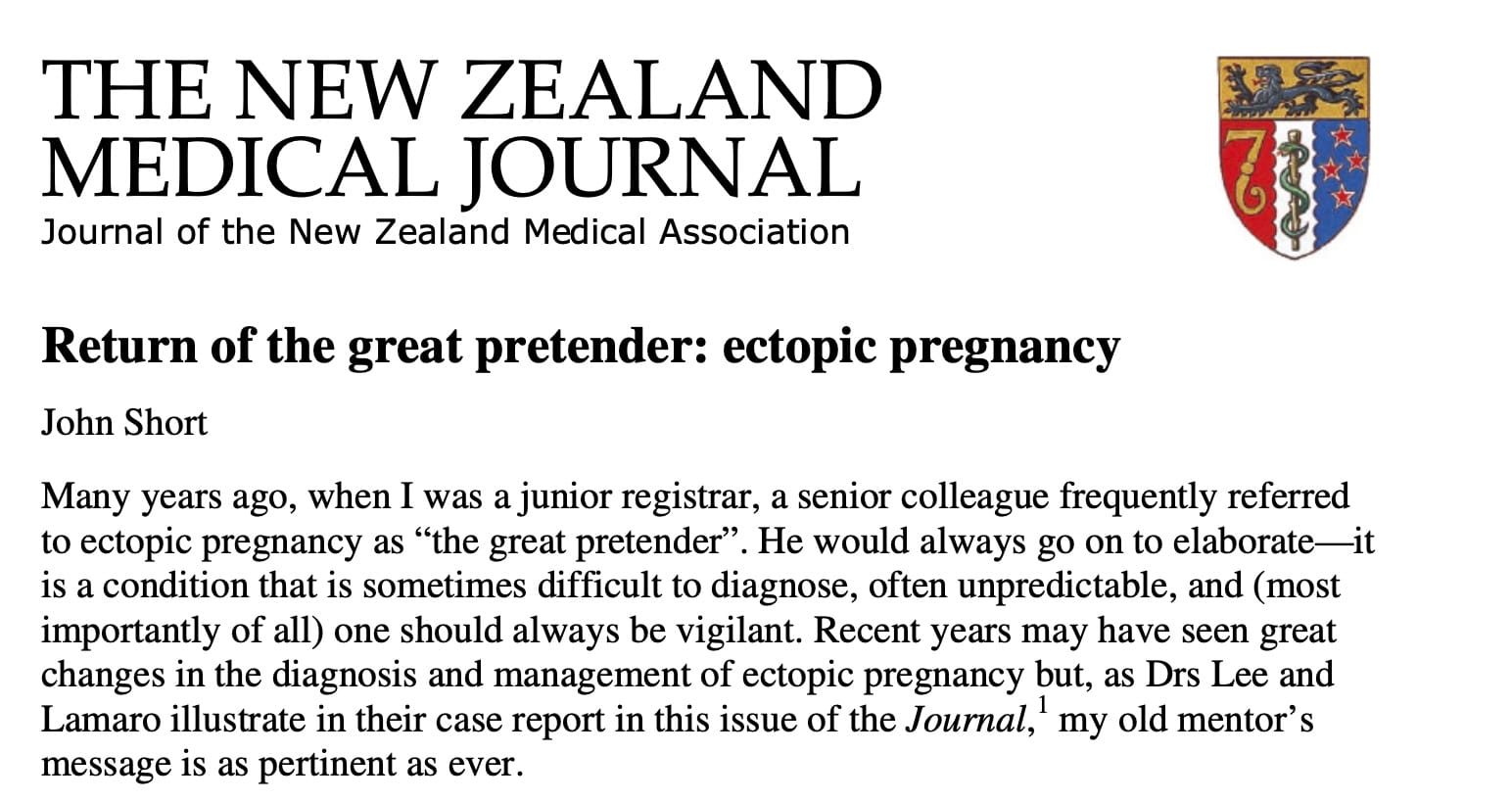
I first came across the term the "great pretender" in relation to ectopic pregnancy in an article in the NZMJ by Obstetric and Gynaecology specialist Dr John Short in 2009.¹ Short explains that "it is a condition that is sometimes difficult to diagnose, often unpredictable, and (most importantly of all) one should always be vigilant". It is a a relatively brief article and although it was written some years ago it is still as relevant as ever. It is one of my all time favourite articles and I know it has saved me from mistakes with ectopic pregnancy.
UK and NZ Data Comparison
Ruptured ectopic pregnancy remains one of the most dangerous—and preventable—emergencies in early pregnancy, and the contrast between the UK and New Zealand is interesting. In the UK, there are approximately 11,000–12,000 ectopic pregnancies each year (around 11 per 1,000 pregnancies or 1:90), with recent national data showing 12 maternal deaths over a two-year period—roughly 6 per year—making ectopic pregnancy the leading cause of death in the first trimester.2 The majority of these deaths are associated with rupture following delayed or missed diagnosis.
In New Zealand, by comparison, there are an estimated 600-700 ectopic pregnancies annually with an incidence —similar to the UK— of approximately 1:100. In the Auckland area there is approximately one ectopic pregnancy for every 64 live births.3
Maternal deaths due to ectopic pregnancy in NZ are thankfully rare. The Perinatal and Maternal Mortality Review Committee(PMMRC) reported 4 maternal deaths from pregnancies with abortive outcomes (ectopic pregnancy and miscarriage combined) between 2006 and 2021.4 Because the PMMRC groups ectopic pregnancy together with miscarriage in its summary tables, the exact number of deaths due solely to ectopic pregnancy is not always reported publicly.
Short estimated that we would expect one death every 5 years in NZ.1 Short also makes the point though that for every death there are likely many near misses—cases of delayed or missed diagnosis which involve substandard care but do not lead to an adverse event or complaint.1 It is difficult to objectively quantify these cases.

The Ectopic Pregnancy Trust (EPT) in the UK has called for urgent action to stop deaths from ectopic pregnancy in light of the 2024 MBRRACE Maternal Deaths and Morbidity Report 2020-2022.2 "During such two-year period in the UK and Ireland, 12 women died from an early pregnancy-related cause. These were all due to ectopic pregnancy. Ectopic pregnancy deaths have risen again – from 5 reported in 2019, to 8 in the 2022 report, to now 12. This is an alarming trend and ectopic pregnancy deaths in this report is almost twice the rate in 2018-20." 2
"The report states that all 12 women who died from an ectopic pregnancy could have had better care. Improvements to care may have made a difference to the outcome for nine women (75%)." 2
The EPT has developed a Think Ectopic Campaign and is working in collaboration with the Royal College of GPs, Royal College of Emergency Medicine and the College of Paramedics to raise awareness of the problem.5
NZ Perspective
The danger in New Zealand with low overall mortality from ectopic pregnancy is that complacency develops amongst clinicians. Low mortality does not necessarily equate to better systems. We know that for any death there are likely many near misses. There are undoubtedly system problems in NZ. Systems of care still need to be continually reviewed and vigilance needs to be maintained. In particular we need to make sure local guidelines reflect current best evidence and are designed to protect patients.
When I was a senior registrar in Emergency Medicine I was asked at one hospital to present some cases of missed ectopic pregnancy at a morbidity and mortality meeting. The department had multiple pathways for the management of a variety of conditions and as registrars we were expected to follow them. I went on to discuss the difficulties of actually diagnosing ectopics and I demonstrated that the department pathway for bleeding in early pregnancy could actually lead to the discharge of a patient with an ectopic. This situation had nearly happened to me the week before. I suggested the pathway be revised. One consultant agreed and complimented me. The senior consultant body including my "supervisor" however suggested that the pathway was fine!
Currently in New Zealand there is immense pressure on frontline medical services. Many patients in urban areas struggle to see their GP in a timely manner. Emergency Departments are overwhelmed. Urgent Care clinics are starting to function as mini community EDs but are under-resourced. There are also many rural communities in NZ which lack ready access to laboratory investigation and ultrasound.
Review of Health and Disability Commission (HDC) complaints in NZ shows other recurring themes—delay in diagnosis despite suggestive features, failure to act on abnormal ultrasound findings, lack of knowledge, poor communication of diagnostic uncertainty, inadequate follow-up planning, premature reassurance and discharge.6,7,8 Much can be gained from reading these HDC reports especially with an appreciation of the evidence-base.
Ectopic Pregnancy: A Lesson in Clinical Thinking
I have no personal interest in gynaecology as a specialty, but what interests me about ectopic pregnancy is what it reveals about how we think as doctors.
Despite a large and long-established evidence base, we continue to make the same mistakes. Ectopic pregnancy is undoubtedly a complex clinical problem, and there are many reasons why errors occur. However, beyond the clinical complexity, there are deeper cognitive and psychological factors that we often fail to recognise and address.
In medicine we make judgements about "likelihood" regarding diagnosis, investigation and management. This process is essential to clinical practice, but it can also mislead us. We should consider the logic—and danger— of trying to make assessments of likelihood when dealing with a highly unpredictable condition like ectopic pregnancy. Many things are considered “unlikely” — until they are not. Unfortunately, in ectopic pregnancy, the consequences of being wrong can be devastating.
Winston Churchill is often quoted as saying: “Men occasionally stumble over the truth, but most pick themselves up and hurry off as if nothing had happened.” There is a lesson here for medicine. We sometimes encounter evidence that challenges our assumptions, but then continue practising as though nothing has changed. We can easily become confused in our thinking and overly confident in our own clinical judgement when we don't keep the evidence-base in mind.
Another challenge is embedded within medical culture. Our training often teaches us to avoid unnecessary escalation. We are encouraged to be measured, rational, and resource-conscious. These are important qualities, but they can have unintended consequences. We may hesitate to refer because we fear being perceived as over-calling a situation. We may avoid the uncomfortable conversation with a gynaecologist colleague. We may choose reassurance over escalation because escalation feels like an admission that we are uncertain.Yet uncertainty is exactly when advocacy is most important.
When advice from a specialist makes us uncomfortable, there can be a tendency to accept it rather than challenge it. The natural human instinct is often to avoid conflict, particularly when speaking with someone perceived as having greater expertise. However, our responsibility is not to avoid disagreement — it is to advocate for the patient.
The essential truth is that safe management of ectopic pregnancy requires three things: clinical humility, evidence-based knowledge, and the courage to advocate for our patients.
If we fail to recognise our cognitive biases, underestimate uncertainty, or allow hierarchy and culture to influence our decisions, we risk repeating the same mistakes. The goal is not to diagnose every ectopic pregnancy perfectly — that is impossible. The goal is to create a system and a mindset where uncertainty is respected, concerns are escalated appropriately, and patients are protected from the consequences of our assumptions
Ectopic Pregnancy Rules
There are several specific recurring diagnostic pitfalls with ectopic pregnancy. One way to counteract this is to make a set of clinical rules which are considered at every decision point in the diagnosis. The evidence base on ectopic pregnancy is very large and sometimes conflicting; it is impossible to do it justice. I include here a brief discussion of some of the evidence but the take home messages are quite simple.
Rule 1. Urine β-hCG in pregnancy is unreliable
False negatives occur due to testing too early, dilute urine, late ovulation or implantation, very low β-hCG levels (<25 IU/ml). Serum testing detects levels as low as 5 IU/L, whereas urine testing detects levels as low as 20–50 IU/L.9
A common problem in daily practice is the young woman with lower abdominal pain with a negative urine β-hCG. Dr. Catherine Varner, Emergency Physician at Sinai Health System describes a case on the EM Cases podcast involving a woman who was initially referred to ED as a possible appendicitis with a negative urine β-hCG. The patient had an elevated serum β-hCG of 52 and went on to be diagnosed with a ruptured ectopic.10
Bottom line : Always do a serum β-hCG on all women of child bearing age with abdominal/pelvic pain or vaginal bleeding regardless of a negative urine β-hCG.
Rule 2. Absence of risk factors does not mean low risk
Approximately 50% or more of patients with ectopic pregnancy have no known risk factors.9,11,12
Bottom line: It is obviously good practice to ask questions regarding risk factors but it should not determine investigation.
Rule 3. Clinical signs and symptoms cannot rule out an ectopic
As doctors we like to take pride in our clinical skill. Unfortunately the clinical diagnosis of ectopic pregnancy is very unreliable.1 The classic symptom triad of abdominal pain, amenorrhoea, and vaginal bleeding is absent in up to 25% of women and up to 10% may have minimal or no symptoms.10
A recurring theme in the literature is delayed diagnosis due to atypical presentations.1 Ectopic pregnancy is a master of disguise. An ectopic pregnancy may masquerade as a miscarriage with isolated vaginal bleeding(light or intermittent), appendicitis or renal colic with unilateral abdominal/flank pain, gastroenteritis with nausea, vomiting or diarrhoea, a urinary tract infection with dysuria or frequency, or even constipation with rectal pressure and pain on defecation— thought to be due to blood in the pouch of Douglas irritating the pelvic peritoneum. Shoulder-tip pain from diaphragmatic irritation is a classic but often overlooked symptom, while some patients present primarily with dizziness or presyncope due to occult intra-abdominal bleeding. Others may have remarkably mild pain despite a ruptured ectopic pregnancy. Significant haemoperitoneum can accumulate gradually.13,14
Pain characteristics are unreliable. Ectopic pregnancy may cause unilateral or bilateral pain, and the severity ranges from mild discomfort to severe pain. Neither the location nor intensity of pain can safely distinguish ectopic pregnancy from other causes of early pregnancy bleeding.Some patients have no pain. 12,14,15
Vaginal bleeding in ectopic pregnancy is typically described as light or spotting, but the amount of bleeding is highly variable. Some women with an ectopic pregnancy experience bleeding that is as heavy as, or even heavier than, a normal menstrual period. The bleeding usually originates from shedding of the decidualised endometrium due to falling progesterone levels rather than from the ectopic pregnancy itself. Studies have consistently shown that the pattern and volume of pain and bleeding are poor discriminators between miscarriage and ectopic pregnancy. Bleeding can be with or without clots.13
Physical examination is of limited value. Some patients have no abdominal tenderness. An adnexal mass is only found in a minority of cases and was present in just 10% of ectopic pregnancies in one study. The absence of an adnexal mass provides is not reassuring.13
The clinical presentation may closely mimic a miscarriage. Some women with ectopic pregnancy even report passage of tissue per vagina, creating the false impression of a complete miscarriage.1,13 Endometrial shedding can also occur with EP.14 It is very easy for patients to mistake blood clot for pregnancy tissue, and accurate differentiation usually requires direct examination by an experienced clinician or by histology. Consequently, the diagnostic value of a patient's report of tissue passage is limited when no specimen is available.13
Assessment of the cervical os is itself imperfect. Determining whether the os is truly open or closed can be difficult. An open external cervical os can be mistaken as an open internal cervical os. If the uterus is antroverted or retroverted, the axis of the cervix and the endometrial cavity lie in different planes making assessment of the os difficult especially for the inexperienced clinician. Furthermore, cervical os status is dynamic and may vary depending on recent passage of blood or clot.13
Bottom line: Symptoms and signs may raise suspicion for ectopic pregnancy, but no combination of symptoms or clinical signs can reliably exclude an ectopic pregnancy.9,10,13,16 Abdominal pain or bleeding in the first trimester should therefore be considered an ectopic until proven otherwise. Any other symptoms in the first trimester should also prompt us to consider ectopic pregnancy.
Rule 4. A β-hCG level cannot rule out an ectopic pregnancy
Ectopic pregnancies can present with highly variable patterns of β-hCG levels—rising, falling, plateauing, or even undetectable.10 The β-hCG pattern of an ectopic can mimic both a normal intrauterine pregnancy and a miscarriage in about 29% of cases.1
A doubling of serum β-hCG levels over 48 hours suggest fetal viability but does not rule out ectopic pregnancy.9 Rising β-hCG levels that fails to reach 50% suggests a failing or ectopic pregnancy, as does a plateau.9 Low or falling β-hCG levels suggest non viability but do not rule out an ectopic nor indicate a benign course of an ectopic.1,9
Ectopic pregnancies do tend to be associated with generally lower β-hCG but the the belief low β-hCG levels (e.g., <1000–1500 IU/L) make ectopic pregnancy unlikely is incorrect.15,17 Several studies have shown a large proportion of diagnosed ectopic pregnancies have β-hCG levels below 2000 IU/L, and documented cases have occurred with levels as low as single digits.
One study showed 50.4% of cases had a β-hCG <1,500 IU/mL, and 8.5% had a level <100 IU/mL.15 The lowest β-hCG level in this study was only 9 IU/mL and this also had ultrasound features.15 Another study found that 41% of ectopic pregnancies had a β-hCG level <2,000 IU/mL at the time of diagnosis, and about 9% had a β-hCG level <100 IU/mL.15
The "discriminatory zone" (DZ)—the serum β-hCG concentration above which an intrauterine pregnancy (IUP) should usually be visible on transvaginal ultrasound— historically 1000-1500 IU/L —can be a misleading. An empty uterus above the DZ raises concern but does not prove an ectopic pregnancy, and a β-hCG below the DZ does not exclude one. Also normal intrauterine pregnancies can have β-hCG levels well above the DZ before a gestational sac is seen. The DZ should be seen as an aid to interpretation—not a diagnostic test. The DZ should therefore be conservatively high such as 3500 IU/ml to avoid aborting a normal intrauterine pregnancy.18 Ultrasound features of ectopic pregnancy may be present at β-hCG levels significantly below the traditional DZ.15
Pregnancy of Unknown Location (PUL) is probably a more useful clinical framework because it acknowledges that, after the initial assessment, there are several possibilities—a very early viable intrauterine pregnancy, a failing intrauterine pregnancy, an ectopic pregnancy or a completed miscarriage.
The PUL approach is superior because (1) it avoids premature diagnosis of ectopic pregnancy(2) it reduces the risk of treating a viable intrauterine pregnancy with methotrexate(3) it encourages structured follow-up with serial β-hCG measurements and repeat transvaginal ultrasound and (4) recognizes that the patient’s symptoms and clinical stability are often more important than a single laboratory value.
Data on PUL varies with some estimates suggesting that 50%–70% are found to have either an EP or miscarriage, and 30% a normal IUP.12 Some data suggests a rise in β-hCG levels less than 35% in 2 days suggests EP with an accuracy of 80.2%.12 A full discussion of the management of PUL is beyond the scope of this article but from the Urgent Care perspective the diagnosis of a PUL situation should mandate immediate referral to a gynaecologist.
Bottom line: There is no single β-hCG value nor pattern of levels that can reliably exclude an ectopic pregnancy.9,10,15 A β-hCG level and should not determine whether ultrasound is performed. Any patient with suspected ectopic pregnancy requires appropriate imaging and clinical assessment regardless of β-hCG level.
Rule 5. A low β-hCG does not rule out rupture or reliably predict a benign course
Ruptured ectopic pregnancies can occur across a wide range of β-hCG values, including very low levels (<100 IU/L). In a retrospective study of 693 ectopic pregnancies 11% of women with a ruptured tube had serum β-hCG levels of less than 100 IU/L.19
In a retrospective case review study of 519 ectopic pregnancies 44% of the patients who presented with evidence of rupture had β-hCG levels less than 1,500 IU/mL The same study showed ruptured and non-ruptured ectopic pregnancies to have similar β-hCG levels.15
There have even been cases hypovolaemic shock in patients with an EP with undetectable β-hCG levels.14
Spontaneous resolution of ectopics can occur through regression or tubal abortion.9 British and Australasian guidelines suggest a β-hCG level of <1500 IU/mL as a criteria for expectant management.17,20 Also they must have minimal pain, no free fluid in the pelvis and be willing and able to attend for follow-up levels.17,20
Success rates vary in the evidence base from 57–100% and seem to relate to the β-hCG level.17 One study reported a success rates of 96% when serum β-hCG levels were less than 175 IU/mL but this fell to 66% if serum β-hCG levels were 175–1500 IU/mL. Other studies have reported similar results of 80–90% if the serum β-hCG levels are less than 1000 IU/mL.17
Tubal rupture is still a possibility and these patients must be carefully followed up until the serum β-hCG concentration falls below 5 IU/mL.3
The β-hCG level does not correlate either size or volume of ectopic.15
Bottom line: β-hCG levels cannot reliably predict the size of an ectopic pregnancy, the presence of rupture, or the risk of rupture.15,19 If any woman who is being managed expectantly presents with increased pain or rising β-hCG level she must be referred to hospital as an emergency.
Rule 6. Normal vital signs do not rule out a ruptured ectopic pregnancy
The dramatic presentation of sudden collapse with hypovolaemic shock from tubal rupture now occurs in fewer than 15 % of cases.11,15 Rupture with haemoperitoneum can occur though with completely normal vital signs. Hypovolaemia, tachycardia, hypotension, diaphoresis, and shock are late signs of rupture.16
A retrospective case review study of 519 ectopic pregnancies found 22.9% presented with evidence of rupture on ultrasound, and 14.4% showed evidence of haemodynamic instability defined as: pulse >100 beats per minute; systolic blood pressure <90 mmHg.15
Multiple studies have demonstrated a poor correlation between vital signs and the volume of haemoperitoneum.22,23 In one study, patients with normal vital signs still had a 20% chance of class IV haemorrhage at surgery.21 Some may even present with relative bradycardia despite losing one to two litres of blood.10,21 This bradycardia is thought to be a vagal response to peritoneal stretching and irritation.
A more useful parameter may be the shock index (SI) or the ratio of heart rate(HR) to systolic blood pressure(SBP). SI may be more sensitive than HR or SBP. Values nearing 1.0 suggest worsening hemodynamic status and shock. A significant elevation in SI (>0.85) may be useful for identifying patients at increased risk of rupture. It should not be used to diagnose or rule out rupture but it could be used as a screening tool.24
Numerous studies and case series have shown that patients can present with significant haemoperitoneum but still appear relatively well. Young patients tend to have greater physiological reserve and can compensate for blood loss but it would also seem that bleeding may occur slowly or intermittently and accumulate without immediate cardiovascular collapse.
An important related question here is wether abdominal examination can exclude haemoperitoneum. Popowski et al 26, amongst other studies, found that abdominal guarding or rebound tenderness independently predicted increasing haemoperitoneum (OR 4.6, 95% CI 2.0–10.8), yet some women with substantial haemoperitoneum lacked these signs, demonstrating that their absence cannot reliably exclude significant bleeding.
There are very few studies in the literature that specifically correlate abdominal examination findings with the measured operative volume of haemoperitoneum. Most papers either describe predictors of tubal rupture or predictors of significant haemoperitoneum (usually ≥300–500 mL) rather than the diagnostic performance of individual examination signs.There are almost no studies that have directly evaluated the negative predictive value of abdominal examination for excluding clinically significant haemoperitoneum in ectopic pregnancy.
This is a subtle but important distinction.The presence of peritoneal signs increases the likelihood of haemoperitoneum, but their absence has never been shown to safely exclude it.
Bottom line: Do not be reassured by normal vital signs. Significant haemoperitoneum and ruptured ectopic pregnancy can be present despite normal blood pressure and heart rate and a benign abdominal examination.
Rule 7. All first trimester patients with pain or bleeding should have a TVS asap
There is no combination of clinical symptoms, physical signs or β-hCG level which can reliably exclude an ectopic.9,10,13 A benign cause for symptoms such as an implantation bleed should never be diagnosed without full investigation. The report of a minor degree of bleeding such as "spotting" does not exclude an ectopic.
All first trimester patients presenting with pelvic pain and/or vaginal bleeding should be investigated for ectopic pregnancy. A transvaginal ultrasound should be performed as soon as possible as the initial investigation and should not be delayed or withheld because of the β-hCG level.9,10,15,18 For many patients this will mean referral to the local ED.
Bottom line: First-trimester pain or bleeding equals ectopic pregnancy until proven otherwise. Arrange a transvaginal ultrasound early, regardless of β-hCG level.
Rule 8. Patients on treatment with methotrexate can still rupture
Patients treated with methotrexate for ectopic pregnancy can still experience treatment failure and tubal rupture. A significant proportion ultimately require surgical management despite initial medical treatment; about 20% of patients in one study required surgical management.15
Any patient receiving methotrexate who develops new or worsening abdominal pain, increasing vaginal bleeding, dizziness, or other concerning symptoms should be assessed urgently for possible rupture.10,15
Bottom line: Methotrexate does not guarantee resolution. Maintain a high index of suspicion for rupture until the ectopic pregnancy has completely resolved.
Rule 9. All patients with potential EP need robust safety-netting and follow up plans
Failure of follow-up is a recurring factor in missed or delayed diagnoses of ectopic pregnancy. Patients lost to follow-up may progress to rupture, resulting in significant morbidity or death. Patients should understand when an ectopic is not fully excluded.
Every patient with a possible ectopic pregnancy should leave with a clear follow-up plan, explicit instructions about when and where to seek urgent care, and an understanding that worsening pain, bleeding, dizziness, syncope, or shoulder-tip pain requires immediate reassessment. 15
Bottom line: Never rely on a single assessment. Clear safety-netting and reliable follow-up are essential components of ectopic pregnancy management.
Rule 10. Think heterotopic in all assisted pregnancies.
Heterotopic pregnancy or the simultaneous occurrence of an intrauterine and ectopic pregnancy has been rare with an incidence of approximately 1 in 30,000 pregnancies. The widespread use of assisted reproduction technologies (ART) such as in vitro fertilization (IVF) has substantially increased its incidence. The incidence of heterotopic pregnancy in IVF recipients is estimated to be 1 in 100.25
Bottom line: Heterotopic pregnancy should be considered in all women with an ART pregnancy, in women with an intrauterine pregnancy with persistent pelvic pain and in women with persistently raised b-hCG levels after a miscarriage or termination of pregnancy.17
References
- Short J. Return of the great pretender: Ectopic pregnancy. NZ Med J. 2009;122:9–12.
Available from: https://www.researchgate.net/publication/23964682_Return_of_the_great_pretender_Ectopic_pregnancy - The Ectopic Pregnancy Trust. The EPT’s statement on the MBRRACE-UK Report 2024. 2024.
Available from: https://ectopic.org.uk/blog/the-epts-statement-on-the-mbrrace-uk-report-2024 - Auckland District Health Board. Ectopic Pregnancy – Diagnosis and Management in Gynaecology and Maternal Fetal Medicine (MFM) Services. 2020.
Available from: https://static.info.content.health.nz/docs/health-pros/topics/maternity/auckland/Ectopic-Pregnancy.pdf - Perinatal and Maternal Mortality Review Committee. Sixteenth PMMRC Report: Perinatal & Maternal Mortality. Health Quality & Safety Commission New Zealand; 2024.
Available from: https://www.hqsc.govt.nz/resources/resource-library/sixteenth-annual-report-of-the-perinatal-and-maternal-mortality-review-committee/ - The Ectopic Pregnancy Trust. Think Ectopic. 2026.
Available from: https://ectopic.org.uk/think-ectopic - Health and Disability Commissioner. Ectopic pregnancy undiagnosed for eight hours. 2024.
Available from: https://www.hdc.org.nz/decisions/search-decisions/2024/22hdc01701/ - Health and Disability Commissioner. Inadequate assessment leading to delayed diagnosis of ectopic pregnancy. 2026.
Available from: https://www.hdc.org.nz/decisions/search-decisions/2026/21hdc01744/ - Health and Disability Commissioner. Management of ectopic pregnancy. 2024.
Available from: https://www.hdc.org.nz/decisions/search-decisions/2024/21hdc00031/ - Murray H, Baakdah H, Bardell T, Tulandi T. Diagnosis and treatment of ectopic pregnancy. CMAJ. 2005;173(8):905–912.
Available from: https://doi.org/10.1503/cmaj.050222 - Helman A. Ectopic Pregnancy Pitfalls in Diagnosis. Emergency Medicine Cases. 2018.
Available from: https://emergencymedicinecases.com/ectopic-pregnancy-pitfalls-diagnosis/ - Schreiber CA, Sonalkar S. Tubal Ectopic Pregnancy. N Engl J Med. 2025;392(8):798–805.
Available from: https://internetbookofemergencymedicine.com/wp-content/uploads/2025/03/tubal-ectopic-pregnancy.pdf - Mullany K, Minneci M, Monjazeb R, Coiado OC. Overview of ectopic pregnancy diagnosis, management, and innovation. Womens Health (Lond). 2023;19:17455057231160349.
Available from: https://www.ncbi.nlm.nih.gov/pmc/articles/PMC10071153/ - Dart RG, Kaplan B, Varaklis K. Predictive value of history and physical examination in patients with suspected ectopic pregnancy. Ann Emerg Med. 1999;33:283–290.
Available from: https://pubmed.ncbi.nlm.nih.gov/10036342/ - Abbott J, Emmans LS, Lowenstein SR. Ectopic pregnancy: Ten common pitfalls in diagnosis. Am J Emerg Med. 1990;8(6):515–522.
Available from: https://linkinghub.elsevier.com/retrieve/pii/073567579090154R - Eisaman DM, Brown NE, Geyer S. Relationship of beta-human chorionic gonadotropin to ectopic pregnancy detection and size. West J Emerg Med. 2024;25(3):431–435.
Available from: https://www.ncbi.nlm.nih.gov/pmc/articles/PMC11112661/ - Crochet JR, Bastian LA, Chireau MV. Does this woman have an ectopic pregnancy? The Rational Clinical Examination Systematic Review. JAMA. 2013;309(16):1722.
Available from: https://emupdates.com/perm/Crochet%202013%20Ectopic%20Rational%20Clinical%20Exam%20JAMA.pdf - Elson CJ, Salim R, Potdar N, Chetty M, Ross JA, Kirk EJ, et al. Diagnosis and Management of Ectopic Pregnancy: Green-top Guideline No. 21. BJOG. 2016;123(13).
Available from: https://doi.org/10.1111/1471-0528.14189 - American College of Obstetricians and Gynecologists. ACOG Practice Bulletin No. 193: Tubal Ectopic Pregnancy. Obstet Gynecol. 2018;131(3):e91–103.
Available from: https://journals.lww.com/00006250-201803000-00046 - Saxon D, Falcone T, Mascha E, Marino T, Yao M, Tulandi T. A study of ruptured tubal ectopic pregnancy. Obstet Gynecol. 1997;90(1):46–49.
Available from: https://journals.lww.com/greenjournal/abstract/1997/07000/a_study_of_ruptured_tubal_ectopic_pregnancy.10.aspx - Royal Australian and New Zealand College of Obstetricians and Gynaecologists. Miscarriage, Recurrent Miscarriage and Ectopic Pregnancy Clinical Guideline. 2025.
Available from: https://ranzcog.edu.au/wp-content/uploads/Miscarriage-Ectopic-Pregnancy.pdf - Hick JL, Rodgerson JD, Heegaard WG, Sterner S. Vital signs fail to correlate with hemoperitoneum from ruptured ectopic pregnancy. Am J Emerg Med. 2001;19(6):488–491.
Available from: https://linkinghub.elsevier.com/retrieve/pii/S0735675701801935 - Pacagnella RC, Souza JP, Durocher J, Perel P, Blum J, Winikoff B, et al. A systematic review of the relationship between blood loss and clinical signs. PLoS One. 2013;8(3):e57594.
Available from: https://doi.org/10.1371/journal.pone.0057594 - Wong W, Tsui K, Tang Y. How useful are vital signs in identifying patients with ruptured ectopic pregnancy? A retrospective correlation study. Hong Kong J Emerg Med. 2011;18(3):155–160.
Available from: https://doi.org/10.1177/102490791101800305 - Koch E, Lovett S, Nghiem T. Shock index in the emergency department: utility and limitations. Open Access Emerg Med. 2019;11:179–199.
Available from: https://www.dovepress.com/shock-index-in-the-emergency-department-utility-and-limitations-peer-reviewed-fulltext-article-OAEM - Maleki A, Khalid N, Patel CR, El-Mahdi E. The rising incidence of heterotopic pregnancy: Current perspectives and associations with in-vitro fertilization. Eur J Obstet Gynecol Reprod Biol. 2021;266:138–144.
Available from: https://doi.org/10.1016/j.ejogrb.2021.09.031 - Popowski T, Huchon C, Toret-Labeeuw F, Chantry AA, Aegerter P, Fauconnier A. Hemoperitoneum assessment in ectopic pregnancy. Int J Gynaecol Obstet. 2012;116(2):97-100. doi:10.1016/j.ijgo.2011.09.019. Available from: https://doi.org/10.1016/j.ijgo.2011.09.019